Imaging Technologies Facility
See what’s really going on inside your samples
Ever wondered exactly how a disease takes hold in a crop? Or why a material behaves the way it does? Sometimes the answer is hiding at a scale the human eye can’t see. That’s where we come in.
The Imaging Technologies Facility at the James Hutton Institute combines advanced microscopes with specialist expertise in sample preparation, imaging and data analysis. Put simply, we help you see the problem clearly, so you can solve it with confidence.
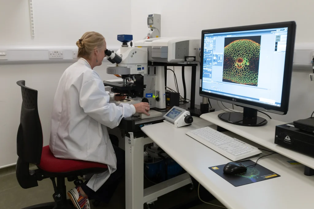
What can we do for you
Think of us as a magnifying glass with a team of experts attached. We can help you:
- Understand plant health and disease. See how pathogens infect plant tissue, how cells respond to stress, and how resistance mechanisms might function.
- Check structure and quality. Examine anatomy, tissue structure and fine detail in biological and material samples.
- Track what’s happening inside living cells. Follow where key proteins sit and how cells react to their environment.
- Obtain publication-ready images and data. Clear, high-resolution results you can use in reports, regulatory submissions, marketing or research papers.
See your challenge clearly
Solve it with confidence.
Who we work with
Wondering whether we’ve worked in your field? Here are some of the sectors and sample types we know well.
-
Plant breeding
Spot resistance, stress tolerance and structural traits at cell level, and back up your selection decisions with hard evidence.
-
Crop protection
Watch how pathogens and pests get into plant tissue, and see how well your treatments stop them.
-
Plant anatomy
Map tissues, cells and fine structure in 3D, from whole organs right down to individual organelles.
-
Agri-food
Look inside grains, fruit and ingredients to check structure and quality, and see what changes during processing.
-
Biotechnology
Track where proteins sit and how living cells respond to new products and processes.
-
Materials
Examine surfaces, coatings, fibres and damage at scales far beyond the reach of a light microscope.
-
Biological samples
Careful preparation and high-resolution imaging for delicate biological specimens.
-
Forensic analysis
Detailed imaging of trace materials, such as soil and plant fragments, to support investigations.
-
Soil and water quality
See the particles, organisms and structure in soil and water samples that routine tests can’t show.
Don’t see yours? Ask anyway. If it fits under a microscope, we’re happy to talk it through.
The kit behind the answers
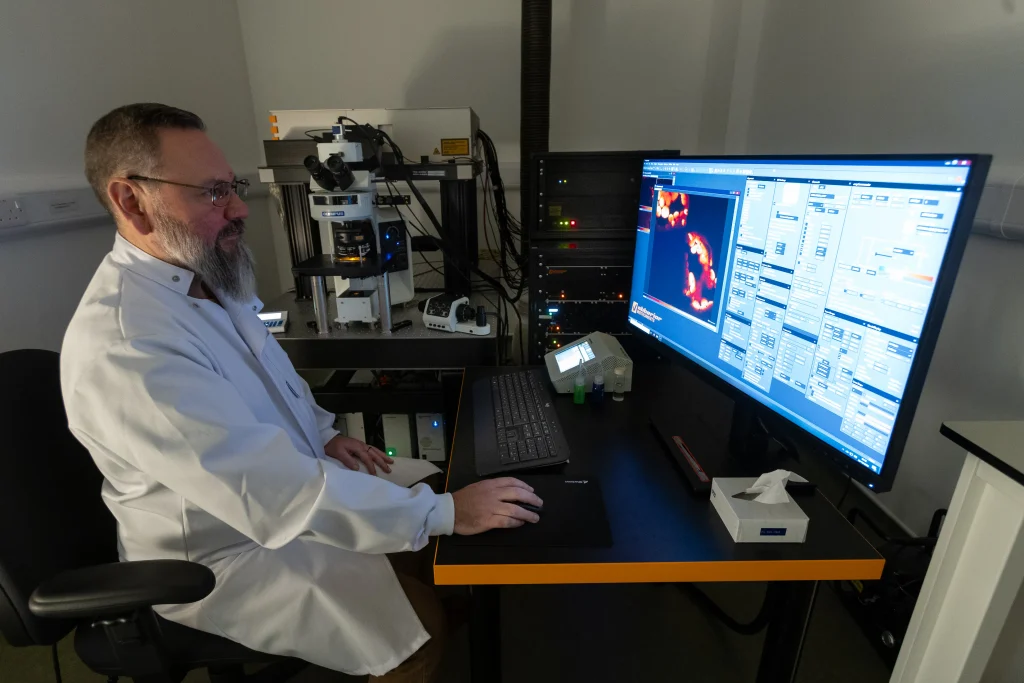
We’ve kept a suite of high-end imaging equipment running for over 20 years. Between them, these instruments cover every scale, from whole tissue down to the tiniest structures.
From sample to answer
Great images start long before anything goes under the lens. Our fully equipped sample processing lab produces high-quality histology samples, including cryosections, wax sections and fresh vibrosliced sections for light microscopy. We can also prepare and section resin-embedded samples for electron microscopy. Our SEM imaging and analysis facility is based in Aberdeen, while all other facilities are based on our Dundee site.
Our experienced team supports you from experimental design right through to data capture, management, analysis and publication. The facility is backed by high-quality data servers, image analysis software and databases. Customers can choose to receive raw data for their own analysis, or we can work together to develop a processing pipeline to deliver publication-ready analysed datasets.
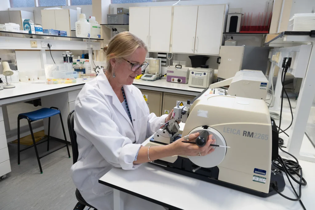
The facility is led by Dr Alison Roberts, Head of Imaging Technologies, supported by senior imaging specialist Dr Michael Porter. Ali and Michael have over 50 years’ experience in microscopy between them and can deliver a wide range of projects. Alternatively, they can work collaboratively to train users to capture their own datasets, so your team builds skills along the way.
Investing in the future
We’ve just secured £640,000 from the BBSRC to install a brand new confocal laser-scanning microscope. It’ll bring higher resolution, faster scanning and better energy efficiency.
We’re doing it sustainably too. Parts of our current system will be reused to create a new widefield microscope, cutting waste and adding another tool to the facility.
Let’s talk about your project
Most of our work happens through scientific collaboration, but we also take on commercial imaging projects. Have a question that needs a closer look? Contact us and we’ll explore what’s possible.
Ramin is your first point of contact for every imaging enquiry, and he’ll bring in Alison and Michael to scope your project.
Contact for more information
Ramin Ebrahimnejad
Business Development Manager
Based in Dundee
Profits from Hutton Scientific Services are reinvested in the James Hutton Institute’s research,
so every project you bring us helps fund science for land, food and climate.